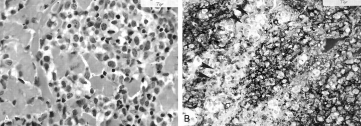
Fig 2.
Case 1. Photomicrographs.
A, Large cells with nuclei display prominent nucleoli that diffusely infiltrate the pituitary gland (hematoxylin-eosin, original magnification ×200).
B, The lymphoma cells are reactive for LCA and stain darkly, in contrast with the normal entrapped pituitary cells (arrowheads) (CD45 immunohistochemical stain, original magnification ×100).

