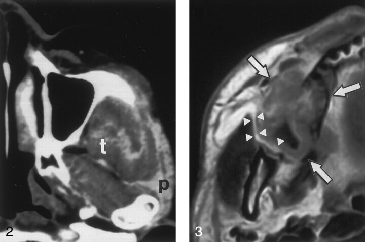
Fig 3.
Axial gadolinium-enhanced T1-weighted MR image in a 45-year-old man with upper gingival cancer in the right molar region shows cancer (arrows), which is spreading into the buccal space but not the masticator space. Note that a thin fat layer (arrowheads) is present between the cancer and mandibular ramus.

