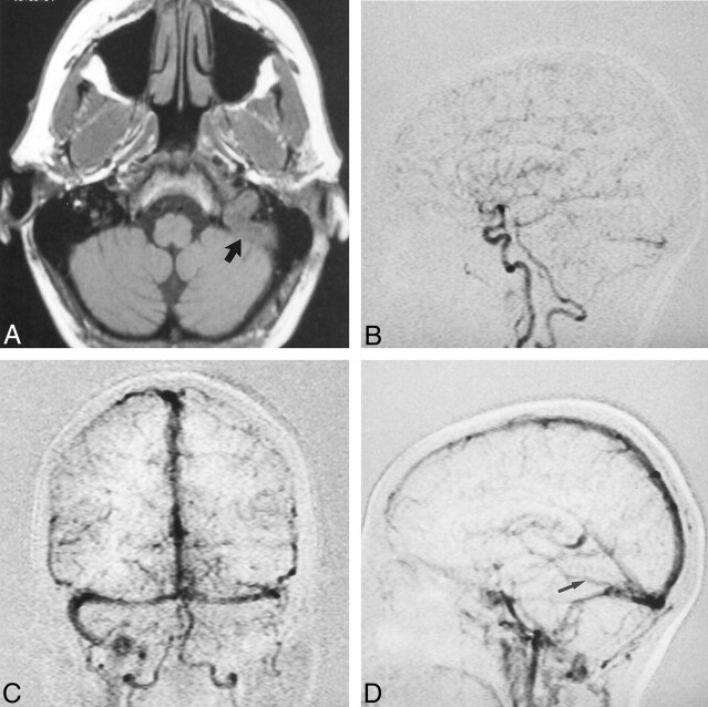
Fig 2.
Case 2. A 51-year-old woman had bilateral tinnitus and an audible bruit over the left mastoid bone.
A, Axial nonenhanced T1-weighted MR image demonstrates marked distension of the left jugular bulb and sigmoid sinus caused by a thrombus (arrow).
B, Single MR-DSA frame from the early arterial phase of the lateral acquisition demonstrates synchronous opacification of the intracranial ICA and left transverse sinus caused by a DAVF fed primarily by branches of the left occipital and posterior auricular arteries.
C, Single MR-DSA frame from the midvenous phase of the frontal acquisition demonstrates enlarged arterial pedicles, occlusion of the left sigmoid sinus, and retrograde venous drainage into the right lateral sinus. Filling of the SSS is not delayed.
D, Single MR-DSA frame from the venous phase of the lateral acquisition demonstrates a large vein of Labbé (arrow) entering the left transverse sinus distal to the DAVF.

