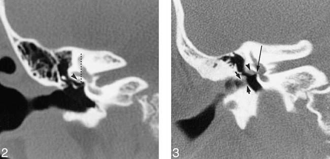
fig 2.

Congenital absence of the oval window, normal facial nerve canal, on coronal CT scan. The oval window is obliterated by a thick bony plate tapering to a central depression. The horizontal facial nerve canal, however, is normal in location (arrowhead), lying lateral to the vertical line drawn through the anterior junction of the lateral and superior semicircular canals.fig 3. Partial absence of the oval window with a large facial nerve on coronal CT scan. This patient with microtia and external canal stenosis (short arrows) has an unusually large horizontal facial nerve (arrowhead). The bone beneath the lateral semicircular canal is thick. The bony facial nerve canal is not well delineated and the nerve overlies the atretic oval window (long arrow)
