
fig 3.
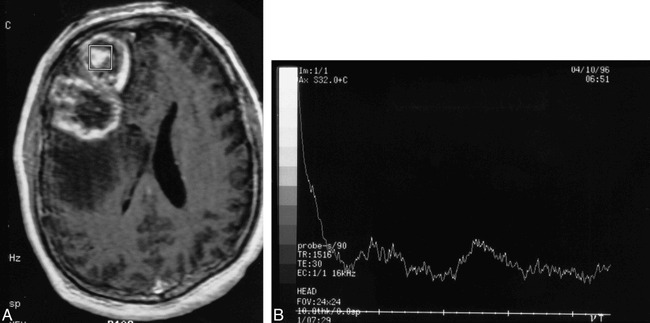
Patient 25: 58-year-old man with a new right frontal lobe mass.
A, Axial T1-weighted MR image reveals a multilobular cystic and solid mass in the right frontal lobe that contains peripheral and central enhancing regions. The spectroscopy voxel is positioned centrally in an enhancing portion of the tumor and does not include the enhancing edge.
B, MR spectrum reveals absence of discernible Cho, Cr, and NAA. The pattern was thought to be consistent with no tumor. The lesion was histologically shown to be a glioblastoma multiforme after resection.