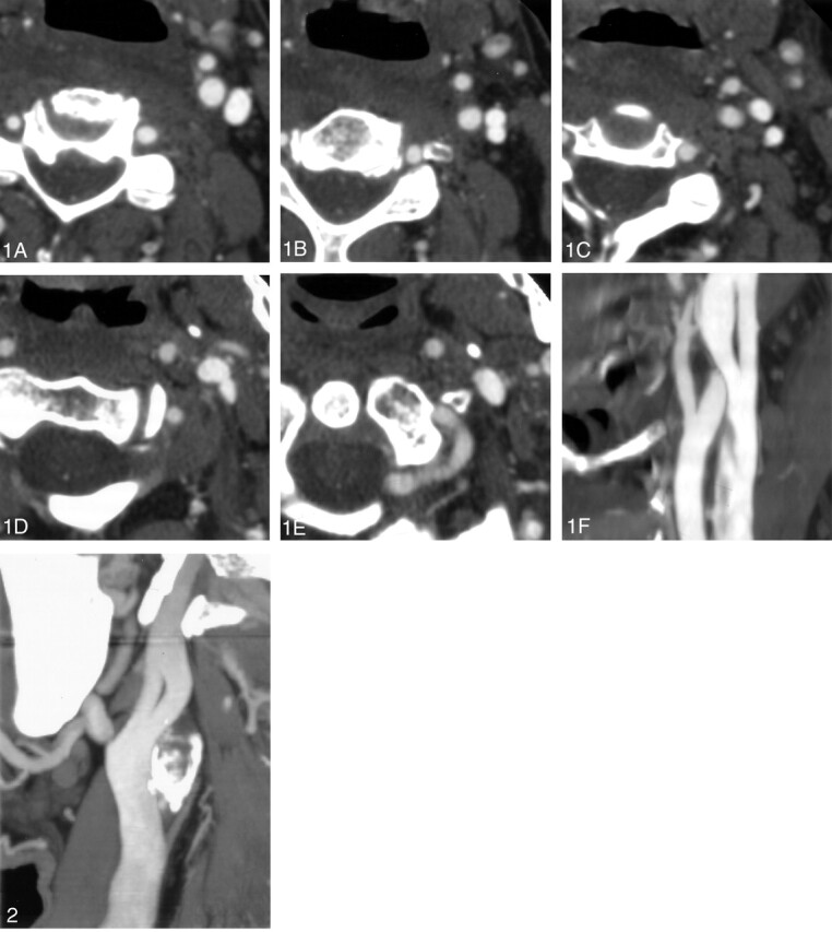
Fig 1.

The appearance of an internal jugular vein on CTA images is characteristic. The vein begins its course with a single lumen (A, B). It then bifurcates forming two distinct lumena (C). As the vein continues caudally, it once again becomes a single vessel (D, E). Panel F shows the fenestrated internal jugular vein reformatted in a single sagittal, oblique plane.
