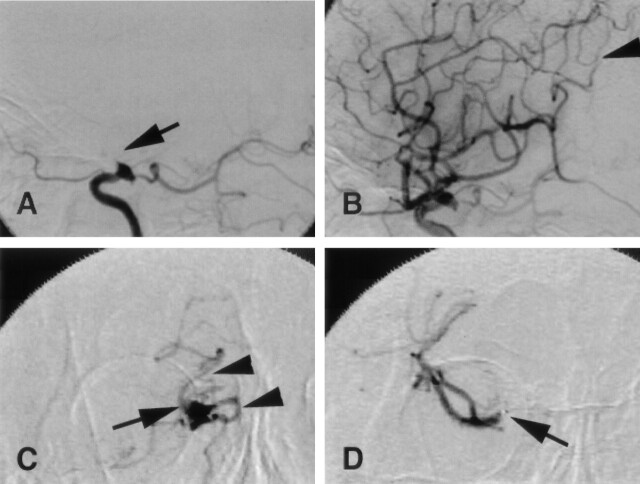
Fig 2.
Case 18. Right internal carotid angiograms, lateral view, before (A) and after (B) thrombolysis and local angiograms from a microcatheter (C and D).
A, Right ICA is occluded distal to the orifice of the posterior communicating artery (arrow).
B, ICA is recanalized. Distal M4 segment of the angular artery is persistently occluded by the fragmented embolus (arrowhead).
C, AP view from a microcatheter proximal to the embolus shows that the ICA is occluded (arrow) distal to the orifice of the posterior communicating artery (arrowheads).
D, AP view from a microcatheter in the distal end of the embolus shows no filling of contrast medium in the proximal MCA M1 segment (arrow). Findings indicate that the embolus extends from the distal ICA to the mid-M1 segment.

