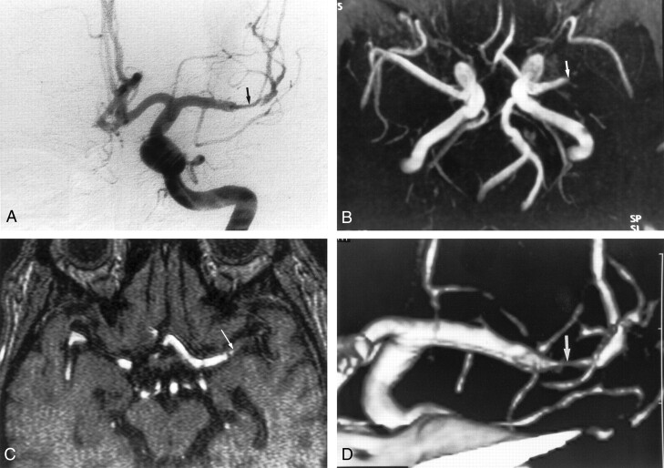
Fig 3.
Images obtained in a 47-year-old man with severe stenosis of the left middle cerebral artery.
A, Anteroposterior angiogram of the left carotid artery shows severe stenosis at the M1 portion of the left middle cerebral artery (arrow).
B, MR angiogram (35/9.6; flip angle, 25°) shows the lesion as an occlusion (arrow).
C, On axial source image (35/9.6; flip angle, 25°), the M1 segment was also interpreted as occlusion (arrow).
D, CT angiogram reveals severe stenosis at the M1 portion (arrow). Two reviewers correctly interpreted this segment as being severely stenotic.

