Fig 2.

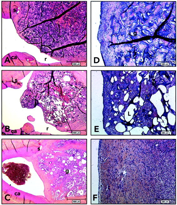
Histologic findings 3 weeks after embolization. ca indicates carotid artery; g, sponge; r, recurrence; S, suture; L, iodized oil. (HPS; magnification ×20 [A–C], ×100 [D–F]).
A and D, Axial section of lateral wall aneurysm and carotid arteries, obtained 3 weeks after embolization with gelatin sponge. Note recurrence and thin neointima covering the uncoated sponge.
B and E, Axial section of lateral wall aneurysm and carotid arteries, obtained 3 weeks after embolization with iodized oil. Note recurrences and thin neointima covering the iodized oil-coated sponges. Higher magnification reveals that the neointima is composed of a few layers of spindle cells embedded in a collagenous matrix and covered by an endothelium in aneurysms treated with iodized oil-coated sponges.
C and F, Axial section of lateral wall aneurysms and carotid arteries, obtained 3 weeks after embolization with cyanoacrylate-coated sponges. Aneurysms treated with cyanoacrylate-coated sponges show a thicker neointima and complete obliteration of the neck.
