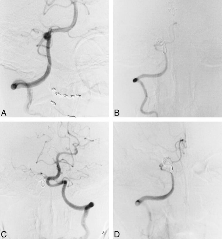
fig 2.

Case 5.
A, Right vertebral artery angiogram, anterolateral view, shows a dissecting aneurysm distal to the PICA origin.
B, Right vertebral angiogram, anterolateral view, immediately after coil embolization of the dissection site.
C, Left vertebral angiogram, anterolateral view, shows an increase in diameter relative to that before embolization.
D, Follow-up right vertebral angiogram, anterolateral view, 1 month after embolization shows complete occlusion of the affected site and preservation of the PICA.
