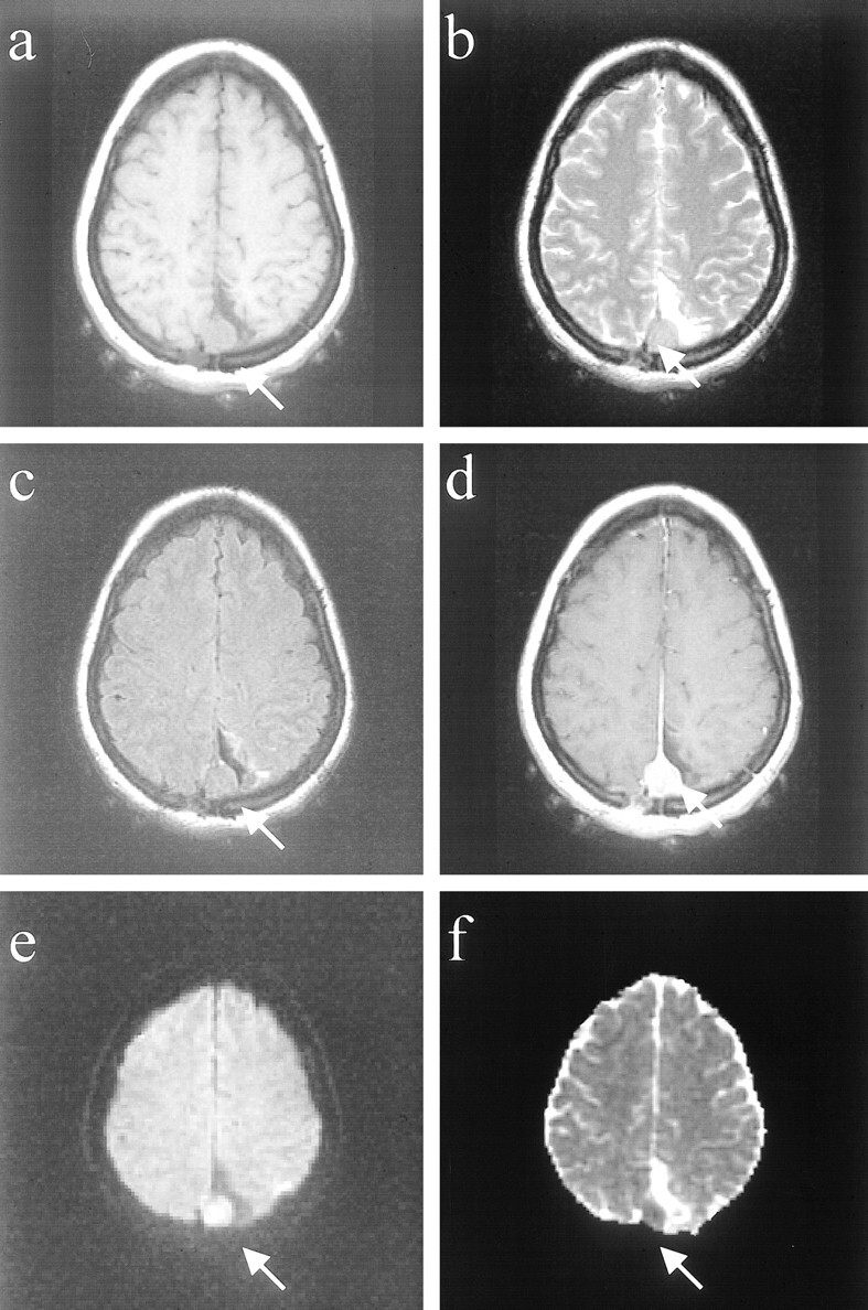
fig 1.

Images of patient (patient 4 in the Table) with a malignant meningioma (WHO grade III).
A, Axial T1-weighted image (500/12/1) shows a well-marginated, recurrent extraaxial mass in the parietal convexity.
B, Axial fast spin-echo T2-weighted image (3000/84/1) of this recurrent meningioma appearing isointense to cortex.
C, Axial fast fluid-attenuated inversion recovery image (10002/162/1) shows isointense mass with smooth borders at resection site and no evidence of brain invasion.
D, Contrast-enhanced spin-echo T1-weighted image (450/12/1) shows smooth, homogeneous enhancement and dural enhancement typical of meningiomas.
E, Diffusion-weighted image (10000/99/1) of the recurrent meningioma, which is hyperintense (lightbulb).
F, Meningioma is hypointense on the ADC map, and the Dav was extremely low (0.45 × 10⁻⁵ cm2/s).
