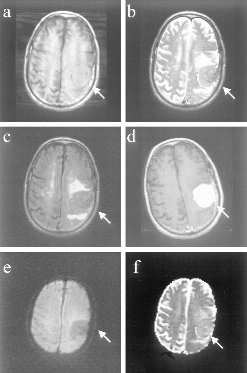
fig 3.

Images of a patient (patient 14 in the Table) with a benign meningioma, distinct histopathologic subtype.
A, Axial T1-weighted image shows left frontal extraaxial mass with smooth margins and pseudocapsule sign.
B, Fast spin-echo T2-weighted image of the mass is isointense to cortex, with areas of hypointensity.
C, Fast fluid-attenuated inversion recovery image shows that the mass is isointense to cortex.
D, Homogeneous enhancement and “dural tail” are seen on the contrast-enhanced axial T1-weighted image.
E, Diffusion-weighted image shows that the meningioma is hypointense.
F, On the ADC map, the lesion is predominantly hyperintense, with areas of isointensity. The diffusion constant of this meningioma was elevated (1.07 × 10⁻⁵ cm2/s). This is greater than a 25% elevation compared with normal brain parenchyma. Histopathologic analysis revealed that this represented a secretory meningioma, which produces brain parenchymal edema (seen best by the fast fluid-attenuated inversion recovery) and which has prominent pericytic proliferations.
