
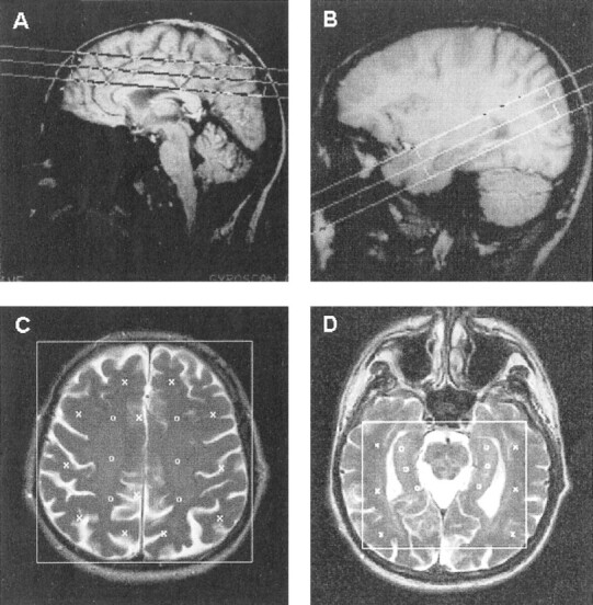
fig 1. MR images identify the position of the PRESS volumes centered at the centrum semiovale and temporal lobe.
A, Sagittal T1-weighted MR image shows the PRESS volume (large box) localized at the centrum semiovale.
B, Sagittal T1-weighted MR image shows the PRESS volume (large box) localized in the temporal lobe.
C, Axial T2-weighted MR image shows the PRESS volume (large box) localized at the centrum semiovale. For metabolite quantitation, 30 voxels were selected and registered on the corresponding MR spectroscopic images. Markers indicate the positions of individual voxels selected in the cortical (x) and semioval (o) regions.
D, Axial T2-weighted MR image shows the PRESS volume (large box) localized in the temporal lobe. For metabolite quantitation, 30 voxels were selected and registered on the corresponding MR spectroscopic images. Markers indicate the positions of individual voxels selected in the temporal (x) and hippocampal (o) regions.