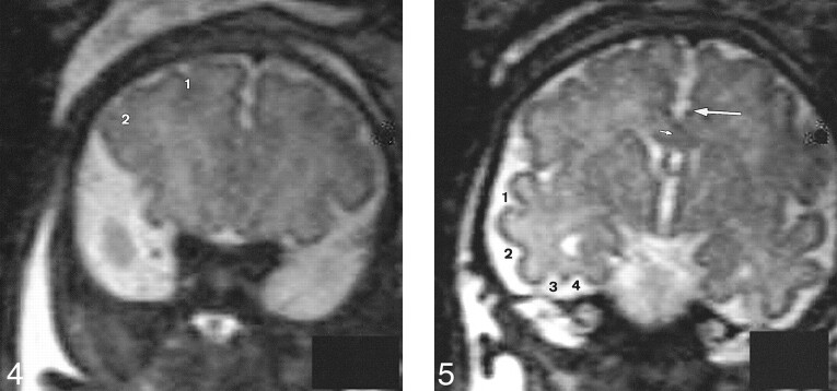
fig 4.
Slice no. 3. T2-weighted anterior coronal image (20/9.2/12) at the anterior part of the frontal lobes (35 weeks' gestation). 1, superior frontal sulcus; 2, inferior frontal sulcus.fig 5. Slice no. 4. T2-weighted coronal image (20/9.2/12) at the level of the third ventricle (35 weeks' gestation). Large arrow, cingulate sulcus; small arrow, callosal sulcus (barely visible); 1, superior temporal sulcus (anterior part); 2, inferior temporal sulcus; 3, external occipital temporal sulcus; 4, collateral sulcus.

