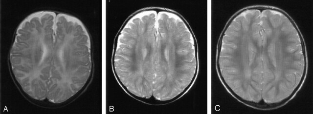
Fig 4.
Patient 2. T2-weighted MR images (3100/98 [TR/TE]). There is a delay in the maturation of the white matter on all three images.
A, At 5 months of age, the unmyelinated periventricular white matter shows no abnormal signal intensities.
B and C, Images obtained at 16 (B) and 35 (C) months of age show nonprogressive, slight signal-intensity abnormalities in the periventricular white matter that mainly involve the occipital trigones.

