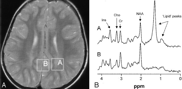
Fig 5.
Patient 3 at 5 years of age.
A, Image shows voxel locations in the occipital trigone (box A) and in the central occipital gray matter (box B).
B, Proton MR spectra (TE = 20 msec) obtained from cerebral white matter (spectrum A) and gray matter (spectrum B). Note the presence of the high, sharp lipid peak at 1.3 ppm and a small peak at 0.8–0.9 ppm in the spectrum obtained from the white matter.

