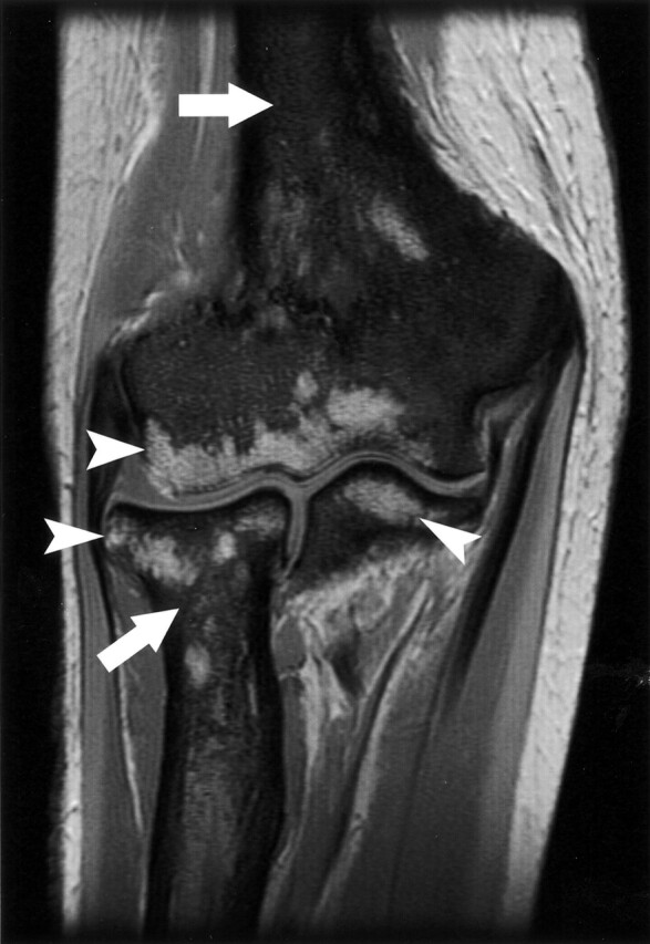
Fig 2.

Coronal T1-weighted image of the elbow demonstrating the replacement of normal bone marrow by low-signal-intensity, diffuse lesions (white arrows) in the humerus, radius, and ulna. High-signal-intensity, normal fatty marrow is spared in portions of the epiphysis of those bones (white arrowheads).
