
Fig 3.
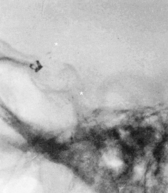
Case 1. Lateral fluoroscopic view of the middle cranial fossa shows a stent in the right ICA and proximal MCA. Stent markers (asterisks) placed proximally and distally enable visualization of the deployed stent. Coils in the supraophthalmic aneurysm are seen between the stent markers in projection on the anterior clinoid process.