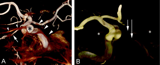
Fig 3.
Volume-rendered 3D-DAs of a right common carotid artery rotational acquisition in 52-year-old woman with a right distal ICA aneurysm, which was incidentally discovered.
A, Lateral view shows the distal right ICA in relation to the adjacent skull. The aneurysm in the superior hypophyseal region (asterisk) and the right ophthalmic artery (arrow) are demonstrated. The relationships of the aneurysm with the clivus (right arrowheads) and the jugum sphenoidale (left arrowheads) are appreciated. Note the specimen-like rendering.
B, Anteroposterior view shows the distal right ICA in relation to the adjacent skull. This view demonstrates the intrasellar location of the aneurysm, which is lying on the floor of the sella (long arrow). Note the jugum sphenoidale (short arrow) and the anterior clinoid processes (asterisk). Also note the dry-bone rendering.

