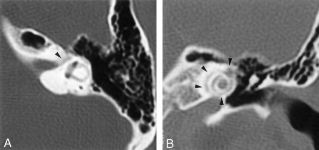
Fig 1.
Initial axial (A) and direct coronal (B) high-resolution CT scans of the temporal bones through the level of the labyrinthine segment of the facial nerve (axial) and the cochlea (coronal) processed with a bone algorithm (2-mm section thickness; 512 × 512 matrix). The facial nerve canal showed slight irregularities along the labyrinthine segment (arrowhead) but otherwise appeared normal. Bandlike, undermineralized areas around the cochlea can be seen (arrowheads).

