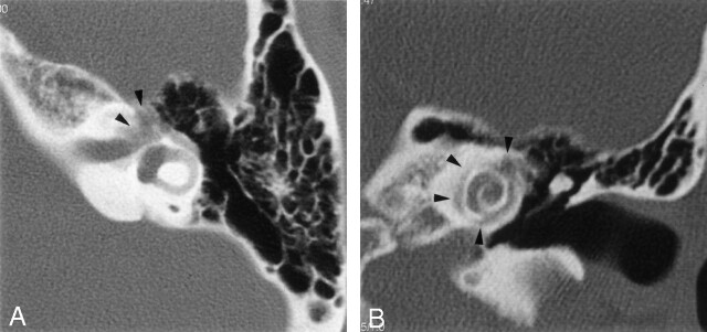
Fig 2.
Further axial (A) and direct coronal (B) high-resolution CT scans through the level of the labyrinthine segment of the facial nerve (axial) and the cochlea (coronal) obtained 2 years after the initial scans (1-mm section thickness; 512 × 512 matrix). The labyrinthine segment, the geniculate ganglion (arrowheads), and the proximal tympanic segment of the facial nerve canal are severely involved and have indistinct, irregular margins. Progression of demineralization is also demonstrated in pericochlear areas (arrowheads).

