
Fig 1.
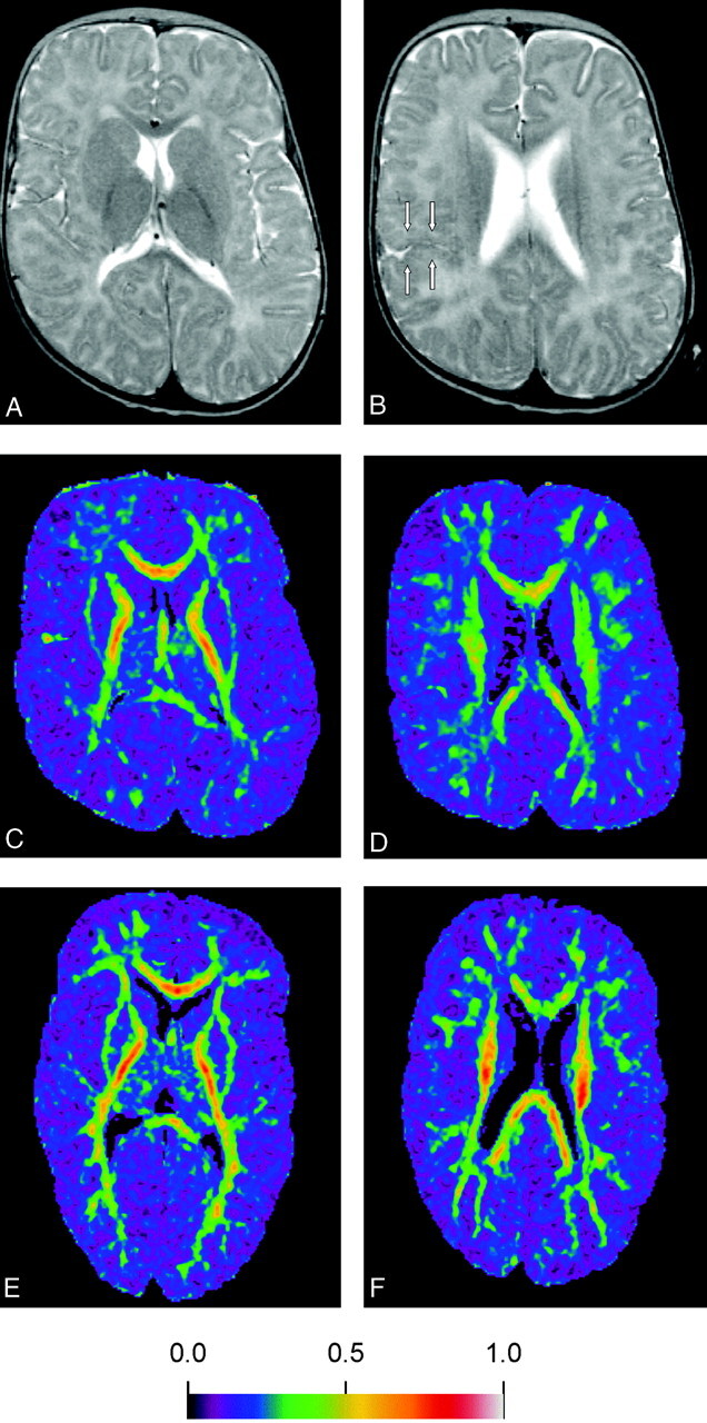
MR images obtained in a 12-month-old boy with biopsy-proved PBD-PTS1 (A-D) and in an age-matched girl with mental retardation (E and F).
A and B, Axial fast spin-echo T2-weighted images (3500/90/1 [TR/TE/NEX]) obtained in a 12-month-old boy, showing diffuse signal intensity increase in the white matter on the level of the anterior limb and genu of the internal capsules and the corona radiata bilaterally, which is consistent with hypomyelination. Posterior limbs of the bilateral internal capsules show hypomyelination. Cortical dysplasia along the border of the right sylvian fissure is present, as can be seen in PBDs (arrows).
C and D, Color-coded FA maps, demonstrating decreased anisotropy in the white matter at the same levels in this patient as those in the control subject (E and F).
E and F, Color-coded FA maps of the age-matched control subject of the corresponding levels as in panels C and D.