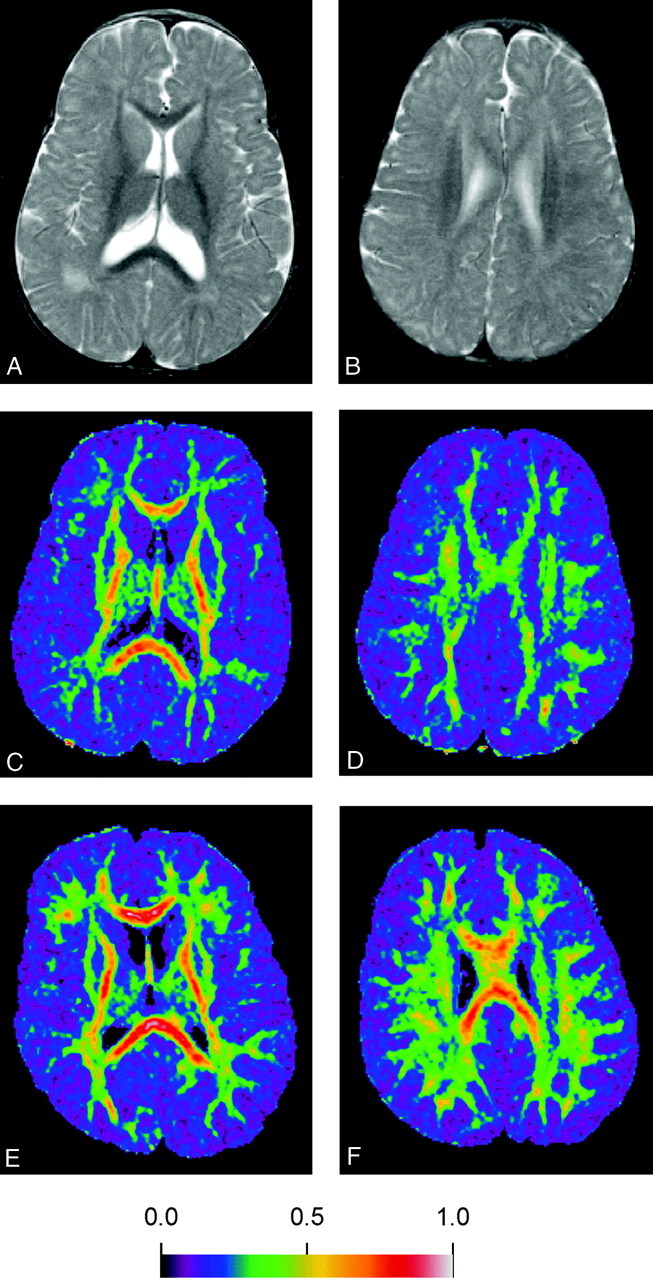
Fig 2.

Images obtained in a 16-month-old girl with biopsy-proved PBD-PTS1 (A–D) and a control subject, an age-matched boy with mental retardation (E and F).
A and B, Axial fast spin-echo T2-weightedimages (3500/90/1), showing high signal intensity in the parietal white matter bilaterally, but normal signal intensity in the bilateral internal capsules, the corpus callosum, and the corona radiata bilaterally.
C and D, Color-coded FA maps, demonstrating decreased anisotropy in the NAWM and abnormal-appearing white matter at the same levels as A and B, as compared with findings in the control subjects (E and F).
E and F, Color-coded FA maps of the age-matched control of the corresponding areas to C and D.
