
Fig 4.
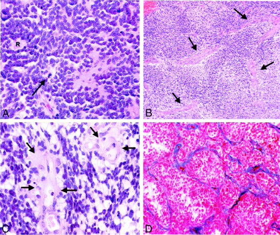
Photomicrographs of PNETs.
A, Hematoxylin and eosin stain shows a hypercellular neoplasm. Poorly differentiated, primitive small cells growing in compact sheets, forming a characteristic Homer-Wright rosette (R), can be seen. Tumor cells have round to oval hyperchromatic nuclei with scant cytoplasm. Mitotic figures are also identified (arrow). The increased cellularity explains iso- or hypointensity of many of the lesions on T2-weighted images (original magnification, ×200).
B, Hematoxylin and eosin stain shows increased vascular channels (arrows) in PNET (original magnification, ×50).
C, Hematoxylin and eosin stain at higher power shows endothelial hyperplasia (arrows) (original magnification, ×400).
D, Azocarmine stain highlights the vascular channels and shows proliferation and thickening of the vascular endothelium within the PNET (original magnification, ×100).