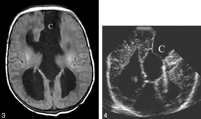
Fig 3.
Neonatal brain MR image taken when the patient was 1 day old. Axial T1-weighted image (similar plane to that of Figure 2B) shows the increased size of the frontal and occipital horns (compare with prenatal scan) in addition to the new finding of a frontal para-midline cyst (C).

