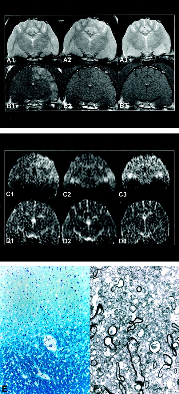
Fig 2.

Images obtained in a cat in group 4: A indicates T2WIs; B, Gd-enhanced T1WIs; C, DWIs; D, ADC maps; E, light photomicrograph; F, electron photomicrograph. 1 indicates 1 hour after embolization; 2, 1 day; and 3, 7 days. At 1 hour, the embolized lesion in the left hemisphere appears hyperintense in A1, enhanced in B1, mildly hyperintense in C1, and isointense in D1. At 1 day, T2WI hyperintensity (A2) and contrast enhancement (B2) are substantially decreased and not evident at day 7 (A3, B3). After day 1, DWIs (C2, C3) and ADC maps (D2, D3) reveal isointensity of the lesion. In E, Light microscopy of the gray matter (top) and white matter (bottom) shows no evidence of demyelination (Luxol fast blue stain, original magnification ×100). In F, Electron microscopy of the gray matter shows no evidence of neuropil or interstitial swelling (original magnification ×3000).
