
Fig 6.
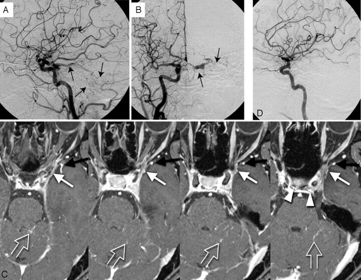
Mixed type with type A and B. A 57-year-old woman with left cavernous dural arteriovenous fistula.
A, Lateral view; B, frontal view. Right carotid arteriogram demonstrates a left cavernous dural arteriovenous fistula draining into the cerebellar cortical veins via the superior petrosal sinus (arrows). Retrograde opacification of the SMCV is not shown.
C, On the contrast-enhanced gradient-echo MR image, a small venous structure connecting to lateral wall of the cavernous sinus, thought to be the SMCV, is observed (white arrow). The findings indicate the potential risk of cortical venous reflux to the medial temporal lobe. Another prominent SMCV flowed into the pterygoid plexus with no connection to the cavernous sinus (black arrow). Note the dilated inferior hypophyseal arteries joining the posteroinferior aspect of the cavernous sinus (arrowheads) and cortical venous reflux to the cerebellar veins (open arrows).
D, This patient was treated by transvenous embolization, and the shunt disappeared completely.