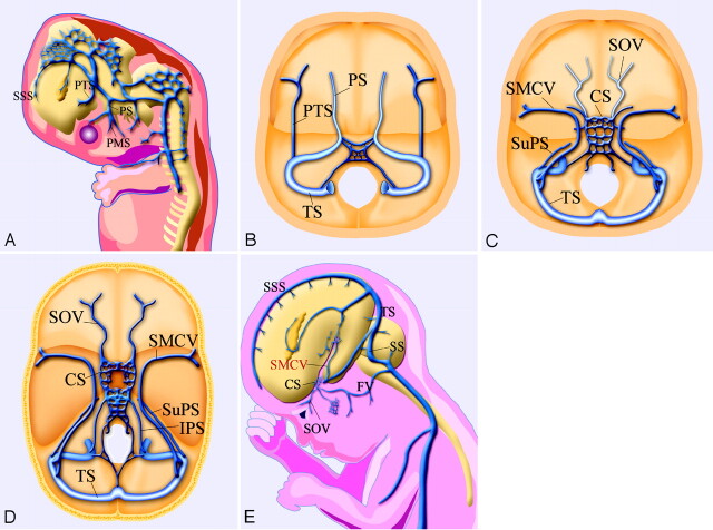
Fig 8.
Schematic drawings of the developmental anatomy of cavernous and para-cavernous venous structures in the embryonic stage. SSS indicates superior sagittal sinus; PTS, primitive tentorial sinus; PS, prootic sinus; PMS, primitive maxillary sinus; and IPS, inferior petrosal sinus.
A, Lateral view; B, axial view. In an 8-week embryo, cerebral venous structures develop from primitive dural plexuses surrounding primitive cerebral vesicles. The plexuses fuse to form venous sinuses and cortical veins. Two major primitive sinuses, the primitive tentorial sinus and the prootic sinus, contribute to the development of cavernous sinus and para-cavernous sinus veins. The primitive SMCV is connected with the transverse sinus via the primitive tentorial sinus.
C, In a 12-week embryo, after several weeks, the primitive SMCV is elongated and develops anteromedially to form the SMCV as a development of the cerebral hemisphere. The prootic sinus contributes to form the superior ophthalmic vein, the cavernous sinus, and the foramen ovale venous plexus.
D, Axial view; E, lateral view. In a developed embryo, the SMCV makes further anteromedial development in the prenatal stage; however, in many patients, there is no direct connection between the SMCV and the cavernous sinus. Secondary anastomosis after birth may form a connection between the SMCV and the cavernous sinus, and the connection to the primitive tentorial sinus subsequently degenerates.

