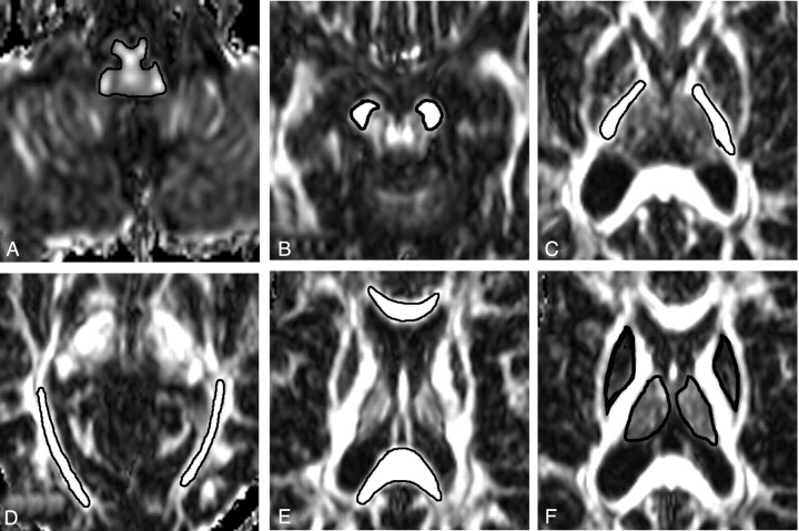
Fig 1.
Examples of region of interest placement for mean diffusivity (MD) and fractional anisotropy (FA) measurements. FA images show regions of interest of medulla oblongata (A), cerebral peduncle (B), internal capsule (C), optic radiation (D), splenium and genu of corpus callosum (E), and putamen and thalamus (F).

