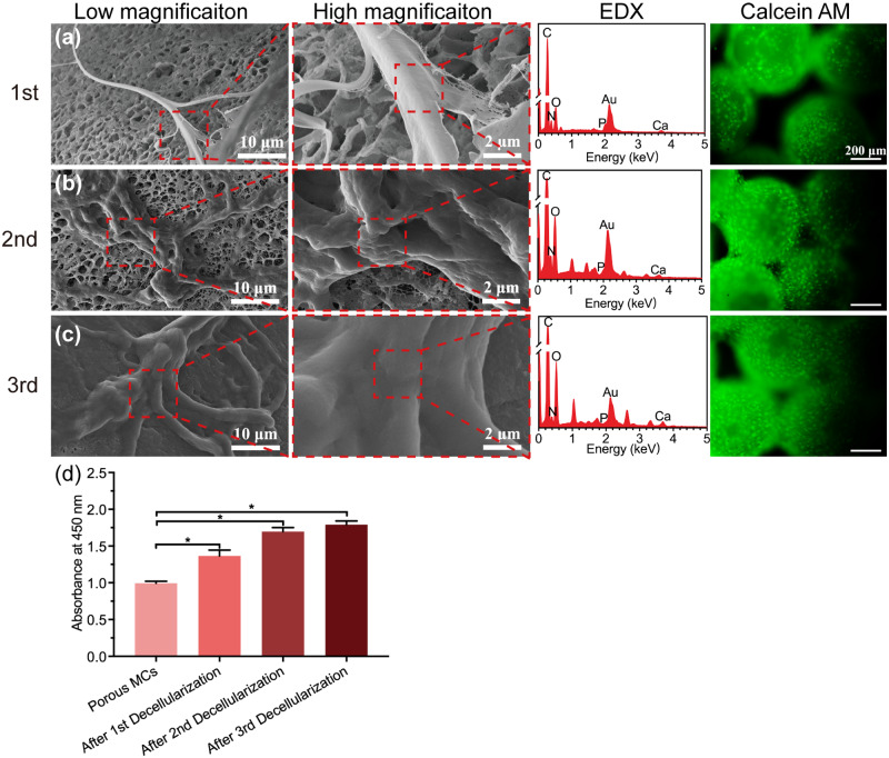
Figure 6.
Characterization and recellularization evaluation of the mECM coated porous microcarriers. The surface microscopic morphology, elemental analysis and cell adhesion of porous microcarriers after first (a), second (b) and third (c) decellularization were evaluated. For cell adhesion assay, the MC3T3-E1 cells were stained with Calcein AM (cytoplasm, green, all scale bar lengths are 200 μm). (d) After 7 days of incubation, the proliferation of MC3T3-E1 cells on porous microcarriers of different decellularization times was evaluated by CCK-8 assay kit (*P ≤ 0.05, n = 3 for each group)

