
Fig 1.
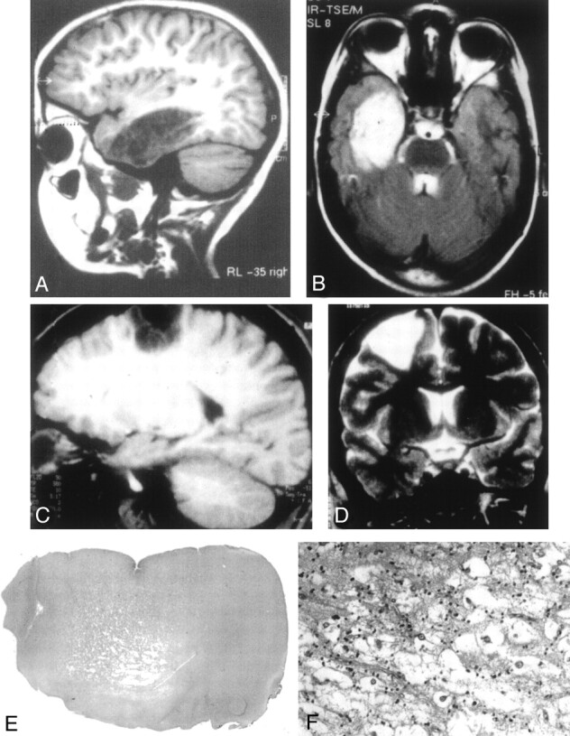
Typical DNT findings.
A, Sagittal T1-weighted MR image shows a large lesion of low signal intensity involving the temporal lobe, without edema or mass effect and corresponding to a complex form of DNT. The lesion is divided by septations leading to an alveolar aspect.
B, The lesion is of high signal intensity on this T2-weighted MR image. The septations appear to be of low signal intensity.
C, Sagittal T1-weighted MR image shows a frontoparietal DNT with sharp boundaries and a rectangular pattern of distribution.
D, Coronal T2-weighted MR image illustrates the triangular pattern of distribution typical of DNT, with a tumor width that is maximal at the cortical level and decreases toward brain ventricles.
E, Low-magnification view showing the cortical location and the nodular architecture typical of DNT (hematoxylin phloxin-saffron, magnification ×10).
F, The glio-neuronal specific element is composed of oligodendrocyte-like cells surrounding areas of mucoid substance containing “floating neurons” (hematoxylin phloxin-saffron, magnification ×300).