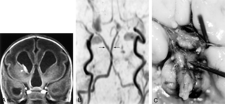
Fig 4.
Images from the case of a premature female patient who was born at 28 weeks (case 4).
A, Coronal view T1-weighted MR image shows duplicated pituitary gland (long arrows). Short arrow indicates germinal matrix bleed on the right side.
B, Time-of-flight MR angiogram shows duplicated distal basilar artery.
C, Autopsy probe points to the origin of the duplicated distal basilar artery. Note the thickened hypothalamus.

