
Fig 3.
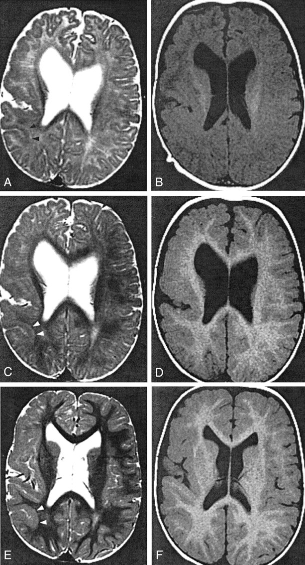
Patient 4, with right hemispheric PMG.
A and B, Images obtained when the patient was 3 months old. T2-weighted image (A) shows pattern 1: 4-mm-thick cortex in the parietal lobe (arrowheads). T1-weighted image (B) shows pattern 2: 5-mm thickness in the same region.
C and D, Images obtained when the patient was 11 months old. T2-weighted image (C) at the parietal lobe shows pattern 1: 4-mm thickness and a 2-mm-thick layer of T2 prolongation between the cortex and myelinated white matter (arrowheads). T1-weighted image (D) shows pattern 2: 6-mm thickness of the same cortex.
E and F, Images obtained when the patient was 2 years old. In the parietal lobe (arrowheads), T2- (E) and T1-weighted (F) images show pattern 2: 6-mm thickness. The T1-weighted image (F) shows pattern 2 in the frontal lobe: 5-mm thickness. Conversely, the T2- weighted image (E) reveals pattern 1 in the frontal lobe: 3-mm thickness with subjacent layer of T2 prolongation.