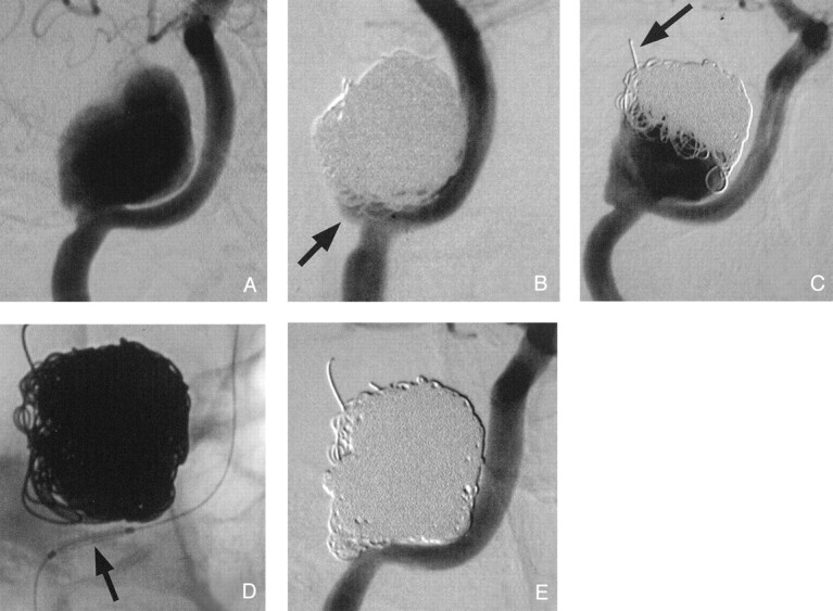
Fig 1.

Endovascular treatment of traumatic ICA pseudoaneurysm.
A, Initial angiogram reveals a large pseudoaneurysm at the carotid canal.
B, After endovascular repair with GDCs, a small aneurysmal neck remnant remains (arrow).
C, GDC packing and aneurysmal growth are noted on the 10-month follow-up angiogram; note coil herniation into middle cranial fossa (arrow). The aneurysmal remnant could not be fully packed with GDCs without the use of endovascular stent reconstruction.
D, As a result, a stent was placed at the aneurysmal orifice (arrow) and was deployed.
E, Additional GDCs could then be detached to complete the repair.
