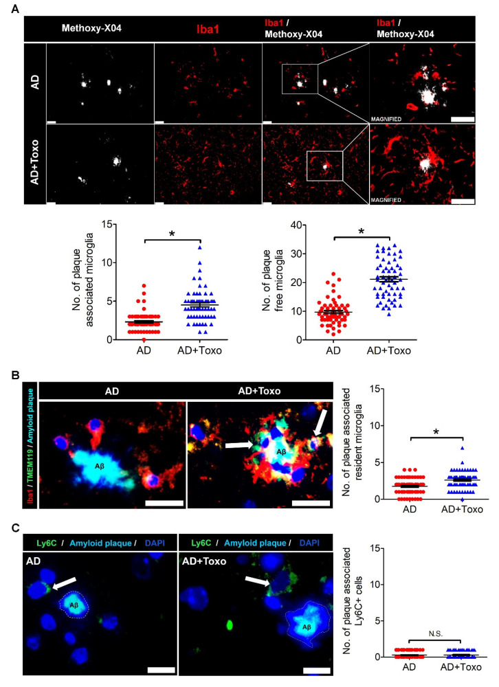
Figure 3.
Plaque-associated patterns of microglia and Ly6C+ monocytes. (A) Iba-1-stained microglia (red) around methoxy-XO4-stained amyloid β (Aβ) plaques (white). The counting result of microglia (plaque-associated or plaque-free) found around 60 amyloid plaques distributed in the brain ((10 randomly selected plaques per mouse) × 6 mice per group). Scale bar; 25 µm. (B) Plaque-associated homeostatic microglia (yellow, due to co-staining of Iba-1 (red) and TMEM119 (green) and indicated by an arrow). Number of plaque-associated homeostatic microglial cells. Scale bar; 20 µm. (C) Ly6C+ monocytes (green) accumulated around Aβ plaque in brain tissues of both AD and AD + Toxo groups. (Ly6C+ (green) monocytes indicated by white arrow). Number of plaque-associated Ly6C+ monocytes. Scale bar; 20 µm. Data are represented as the mean ± SEM. * Statistical significance compared with the control (* p < 0.05).

