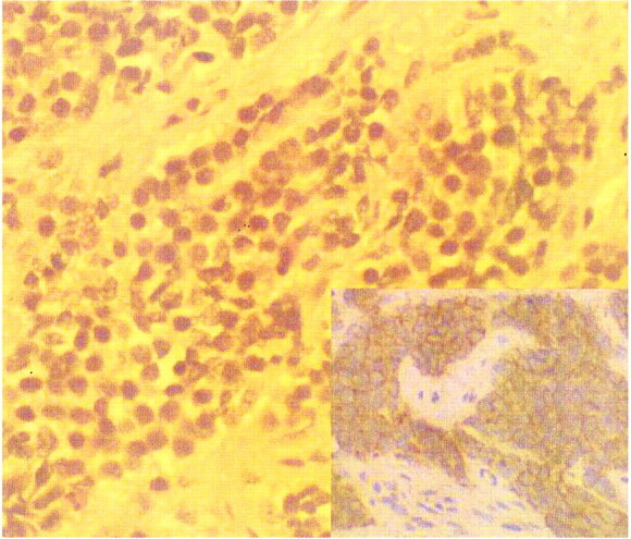
fig 2.

Case 1. Photomicrograph (hematoxylin-eosin stain, magnification ×400) shows nests of small, uniform round cells in the fibrous stroma. Inset: Diffuse intense membrane reactivity for CD99 (MIC 2) on immunohistochemical stain (magnification ×200), consistent with extraskeletal Ewing sarcoma
