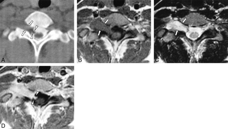
fig 3.

Case 2: 22-year-old woman with numbness of right forearm.
A, CT myelography shows an epidural mass displacing the thecal sac to the left side and intradural filling defects (arrowheads), representing the intradural component at the C7–T1 level. Ipsilateral neural foraminal widening is evident (arrows).
B and C, T1-weighted (B, 500/12/2) and T2-weighted (C, 3200/96/2) axial MR images show a well-demarcated epidural mass at the C7–T1 level, which is isointense to muscle with T1 weighting (B) and hyperintense to muscles with T2 weighting (C). Ipsilateral neural foraminal widening is also observed (arrows).
D, Contrast-enhanced T1-weighted (500/12/2) axial MR image reveals homogeneous, moderate enhancement. The enhancing intradural component connected to the epidural mass is clearly visible within the right side of the thecal sac (arrowheads).
