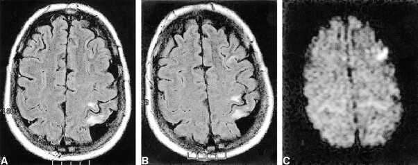
fig 1.
Images show interval appearance of signal hyperintensity in the left middle frontal gyrus.
A, Axial fluid-attenuated inversion recovery image obtained 15 days before surgery.
B, Axial fluid-attenuated inversion recovery image obtained 1 day after left carotid endarterectomy.
C, This finding is hyperintense on the diffusion-weighted image, consistent with acute ischemia. Note the lack of bright diffusion signal in the chronic perirolandic infarct.

