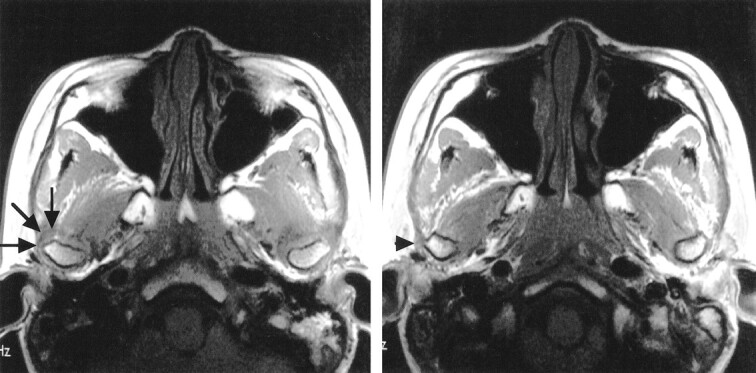
fig 4.

Same patient as shown in figure 3. A series of continuous axial T1-weighted images at the level of the TMJ obtained while the patient was in the closed-mouth position. Note a part of anterolaterally displaced disk (arrows) covering the lateral surface of the right mandibular condyle and a part of widened articular capsule (arrowhead) adjacent to the lateral surface of the condyle
