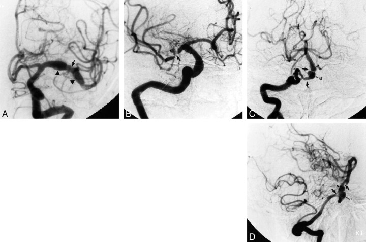
fig 2.
Cerebral angiographic images obtained in 1997.
A and B, An anteroposterior projection of the left internal carotid artery (A) and a 20° left posterior oblique projection (B) of the right internal carotid artery also reveal alternating areas of stenosis (arrow) and ectasia (arrowheads). Note stenotic areas in the course of the anterior temporal artery. No evidence of aneurysm was noted in the internal carotid artery distributions.
C and D, Anteroposterior projection (C) and lateral projection (D) of the selected injection of the right vertebral artery show alternating areas of ectasia (arrowheads) and stenosis (arrow) involving the distal segment of the right vertebral artery and the proximal segment of the basilar artery. A small aneurysm (a) is present at the vertebrobasilar junction.

