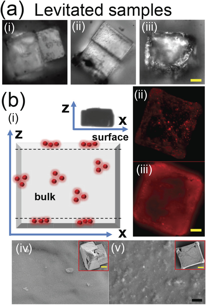
FIG. 3.
Micrograph of the preserved precipitate from levitated droplets of (i) φnp = 0, (ii) φnp = 0.1, and (iii) φnp = 0.0001 (viral load) and (b) (i) the schematic of the levitated precipitate with viral load showing entrapped nanoparticles (red spheres). The symbol z represents the levitator axis while x represents the corresponding perpendicular direction (inset shows the final shape of the levitated precipitate). (ii) The fluorescent image of the precipitate at depths of (ii) z = 1 µm (on the surface) and (iii) 21 µm (within the bulk). (iv) SEM of φnp = 0 (inset shows the complete precipitate under SEM). (v) The same sequence of images as (iv) for φnp = 0.0001. Scale bars in yellow equal 20 μm and in black equal 1 µm.

