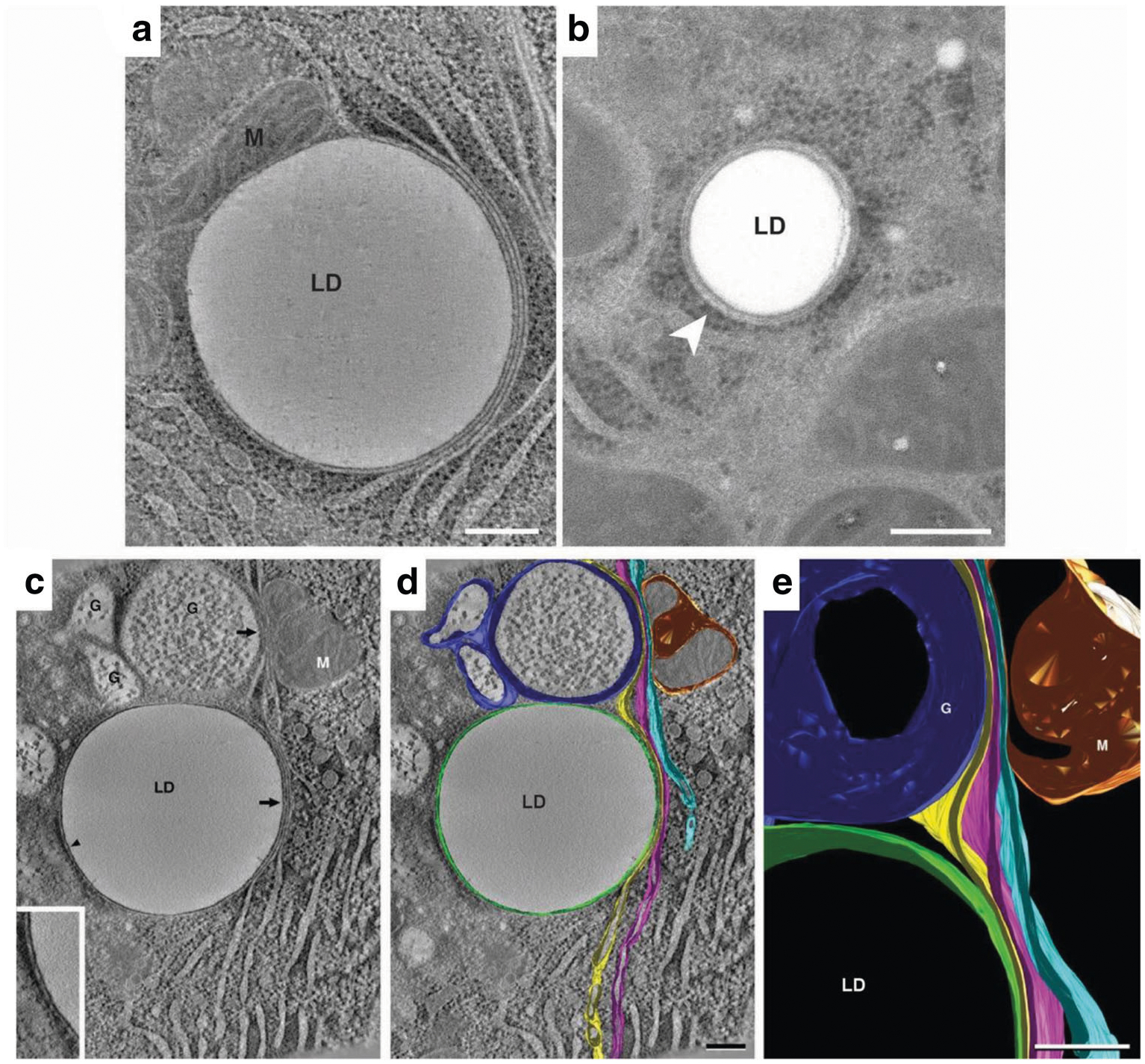
Fig. 2.

Endoplasmic reticulum association with lipid droplets in mammary epithelial cells on lactation day 10. (A) A representative composite of 15, 0.9 nm slices from a tomographic reconstruction of a MEC from an L10 mammary gland prepared by high-pressure freezing and freeze substitution. Domains of three distinct ER cisternae exist in tight association with the lipid droplet (LD). One cisterna wraps around ~60% of the droplet’s perimeter. The other two are wrapped tightly adjacent to the first cisternae on the right side of the droplet. All three cisternae extend beyond the droplet into the cytosol. Ribosomes are absent from ER domains that contact the LD surface or other cisternae but are present on domains facing the cytosol. A closely associated mitochondrion (M) is present on the left side of the LD. (B) Thin (40 nm) section of a LD in a rat hepatocyte. The droplet is smaller than those in L10 MEC, but it associates with two ER cisternae in a similar fashion and only the cytoplasmic face of the outer ER cisterna bears ribosomes. Bars = 0.2 μm. (C-E) Tomographic reconstruction of 10, 0.9 nm slices from another L10 MEC. (C) A tomographic slice shows a lipid droplet (LD) in contact (arrowhead) with single ER cisterna (inset), which is juxtaposed to three additional slender ER cisternae to form a four-layer domain that is in contour with a portion of the LD surface (arrow). The outer cisternae extend upwards from the lipid droplet to approach both the trans-region of a nearby Golgi complex (G) and a mitochondrion (M). (D) The same image data shown in panel C overlaid with filled model contours of ER cisterna, Golgi and Mitochondria. The cisterna in contact with the LD is shown in green, the three juxtaposed cisternae are shown in yellow, magenta, and light blue respectively. The yellow and light blue ER cisternae form similarly close appositions with the trans-Golgi (dark blue) and mitochondrion (gold) respectively, with the magenta cisterna between them. Domains of ER cisternae in contact with each other or with the other organelles are free of ribosomes but bear ribosomes on cisternal regions that face the cytoplasm. (C) Detail of the projected model showing specialized ER domains in close apposition to the lipid droplet, Golgi (G), and mitochondrion (M). Bars = 0.2 μm
