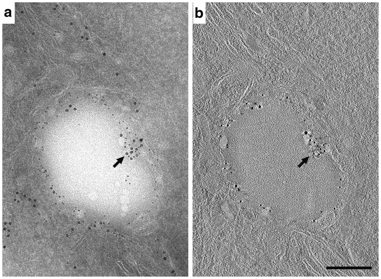
Fig. 4.

Immuno-EM localization and tomographic reconstruction of Plin2 on lipid droplets in HEK293 cells. (A) A 100 nm section of an HEK293 cell double-labeled with antibodies to PDI (15 nm gold) and Plin2 (10 nm gold). PDI localizes to regions of ER that are proximal and distal to the LD surface. Plin2 localizes to the LD surface. A portion of the ER that intercalates into the right side of the LD is labeled with antibodies against both PDI and Plin2 (arrow). (The inset shows an enlargement of this area). (B) A single 0.77 nm slice taken from the middle of the interior of the tomographic reconstruction shown in (A). Plin2 labeling is present through the interior of the section and the co-labeled region (arrow) is easily distinguished, indicating that it is indeed an intercalation and not a superimposition of a labeled structure above or below the lipid droplet (The inset shows an enlargement of this area documenting the presence of both immunogold labeled Plin2 and PDI). Bar = 0.2 μm
