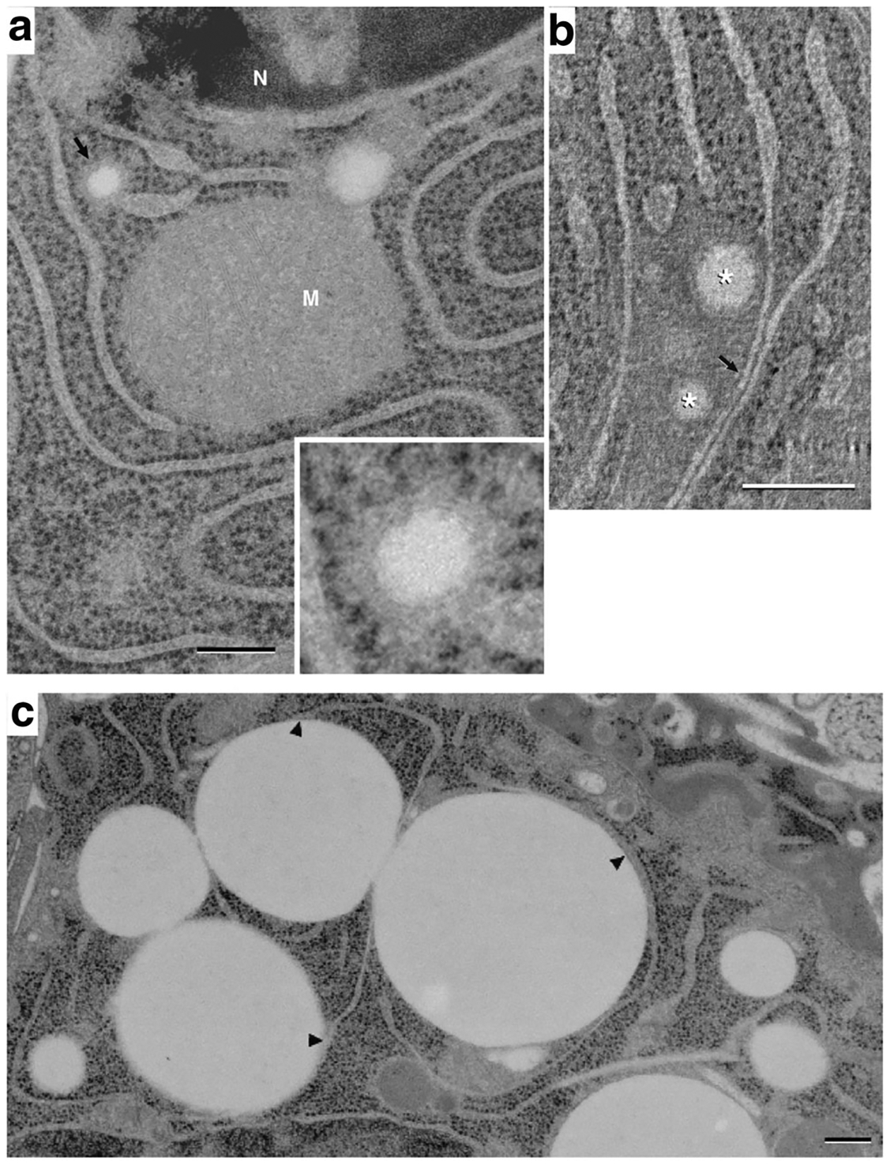
Fig. 5.

Endoplasmic reticulum association with lipid droplets in differentiating mammary epithelial cells. (A) Thin (40 nm) section image of a mammary epithelial cell on pregnancy day 10 (P10), showing a small (~100 nm) lipid droplet within the lumen of an ER cisterna (arrow and inset). The small droplet is surrounded by a dense material that is similar in appearance to, and is continuous with, the ER lumen. This density is itself surrounded by a ribosome-bearing membrane that is continuous with the ER membrane. A second, slightly larger lipid droplet is present on the upper right side of the mitochondrion (M). Although it is surrounded by dense material similar to that of the smaller droplet, its association with nearby ER cannot be determined in the plane of this thin section (N, nucleus). (B) A composite of 7, 1.1 nm slices from a tomographic reconstruction of a P10 cell, showing 2 small lipid droplets (*) amongst ER cisternae. The droplets lie within a matrix that excludes ribosomes; regions of ER cisternae bordering the matrix are smooth. Two ER cisternae near the droplets are closely apposed to one another analogously to the ER domains associating with larger droplets (arrow). Bars = 0.2 μm. (C) EM image showing a field of LD in contact with ER cisternae in a mammary epithelial cell at P5. ER cisternae are shown to contact multiple mature (1 μm) droplets at numerous points (arrowheads) while other parts of the reticulum remain distant from the droplets. Small (~100 nm) LD localize to termini of ER cisternae (asterisks)
