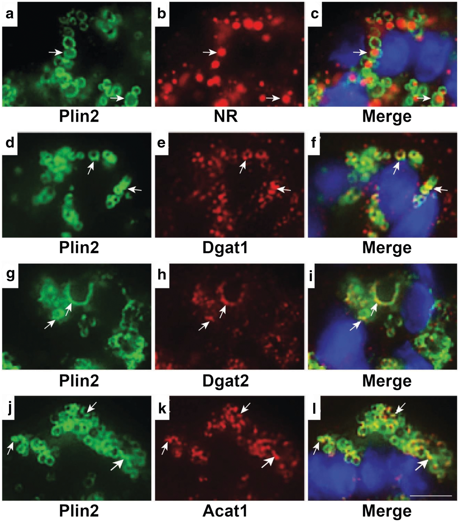
Fig. 6.

Localization of ER proteins and lipid droplets in mammary acini of pregnant rats. Representative confocal microscopy images of mammary acini from day 5 pregnant rats showing specific localization of Plin2 to the surface of Nile red (NR) stained LD (arrows, A-C). Areas of overlap of Dgat1 (arrows, D-F); Dgat2(arrows, G-I); and Acat1 (arrows, J-L) with Plin2 indicate their localization on the LD surface. DAPI-stained nuclei are shown in blue. Bar = 7 μm
