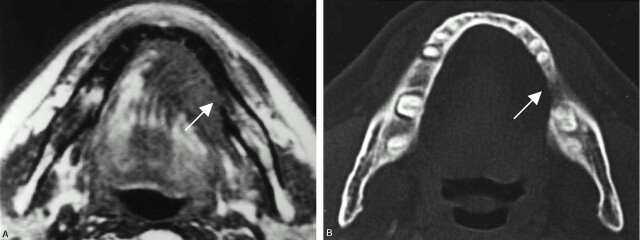
Fig 1.
A 67-year-old man with left floor-of-mouth carcinoma, showing a true-positive result for cortical invasion with both MR imaging and CT.
A, Axial T1-weighted MR image (560/14).
B, Axial bone algorithm CT image.
Both MR and CT image reveal destruction of the cortex (arrows) adjacent to the tumor mass. These findings were histopathologically confirmed.

