Fig 4.
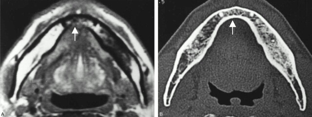
A 46-year-old man with anterior floor-of mouth carcinoma showing a false-positive result with MR imaging and true-negative result with CT for cortical invasion.
A, Axial T1-weighted MR image (560/14).
B, Axial bone algorithm CT image.
The lingual cortex (arrow) is suspected to be involved by the tumor mass on T1-weighted MR image, whereas it is intact on CT. Histopathologic examination after marginal mandibulectomy confirmed no tumor invasion into the mandible. As in Fig 3, misevaluation with MR imaging is considered to be due to chemical shift artifacts.

