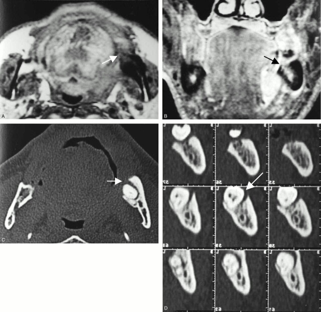
Fig 5.
A 62-year-old man with left floor-of mouth carcinoma showing a false-positive result with MR imaging and true-negative result with CT for cortical invasion.
A, Axial T1-weighted image (560/14).
B, Contrast-enhanced coronal T1-weighted image (700/14).
C, Axial bone algorithm CT image.
D, Dental CT reformatted images.
MR images reveal diffuse abnormal signal intensity of the bone marrow in the left molar region (A and B, arrow), strongly suggestive of tumor invasion. On the other hand, CT reveals bone absorption with relatively smooth margins around the tooth root (C and D, arrow), suggestive of periodontal disease. Histopathogic examination after marginal mandibulectomy confirmed no tumor invasion into the mandible.

