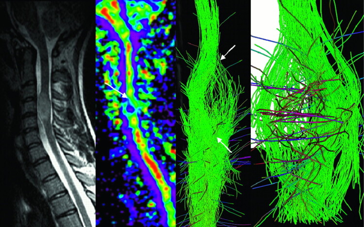
Fig 2.

MR imaging of a spinal cord involvement due to a solid state astrocytoma. FA map and fiber tracking over b0 image show warped fibers around the tumor. Neither the vasogenic edema nor the cystic portion of the tumor was visible on the T2-weighted image, and boundaries of the lesion visible on both the FA map and 3D FT reconstructions (arrow) matched those on T2-weighted imaging.
