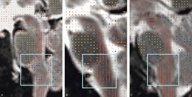
Fig 1.
A, Diffusion tensors overlaid on T2 image in the patient with cervical trauma. Disorganization of the distal brain stem white matter tracts is present.
B, Diffusion tensors overlaid on T2 image in the patient who died after cardiac arrest shows intact fibers with an overall slight wavy configuration.
C, Diffusion tensors overlaid on T2 image in a healthy living volunteer showing the homogenous character of the craniocaudal brain stem white matter tracts.
The principal eigenvector of the diffusion tensor scaled by the fractional anisotropy (FA) is represented by its projection onto the sagittal plane by using red dashes and by its through-plane component by using dots (yellow dot for strong component, blue dot for intermediate component, and missing dot for a weak component). For clarity, tensors were visualized in this figure at a lower resolution than measured, displaying a single vector for every 9 pixels of the interpolated image (ie, 1.5-times lower resolution in both dimensions than measured).

